Breed: Württemberger Warmblood
Gender: Gelding
Age: 14 years old
Discipline: Allround
Comet was purchased two years ago without a PPE. He presented as acutely lame in the left front (LF) after shoeing — “hot nail”. However, after the hot nail healed, Comet was still lame and Dr. Michael Schöberl was asked to assess the horse and provide a second opinion. Schöberl is an ISELP certified veterinarian.
The first examination was performed on February 17.
Hoof tester: negative
All flexion tests negative, except for:
*All lameness grades presented in this case study refer to the AAEP scale.
Analysis revealed a moderate LF asymmetry on both the straight line and on the longe. Following treatment of the shoulder and coffin joint, the horse was sound for a time. The analyses in this case study were performed at a fourth re-check on October 22, and illustrate the use of data to evaluate the effect of diagnostic analgesia.
Examination surface: Hard
As the trot-up lane available was quite short, Comet was trotted away from the camera and back one extra time, three times instead of twice.
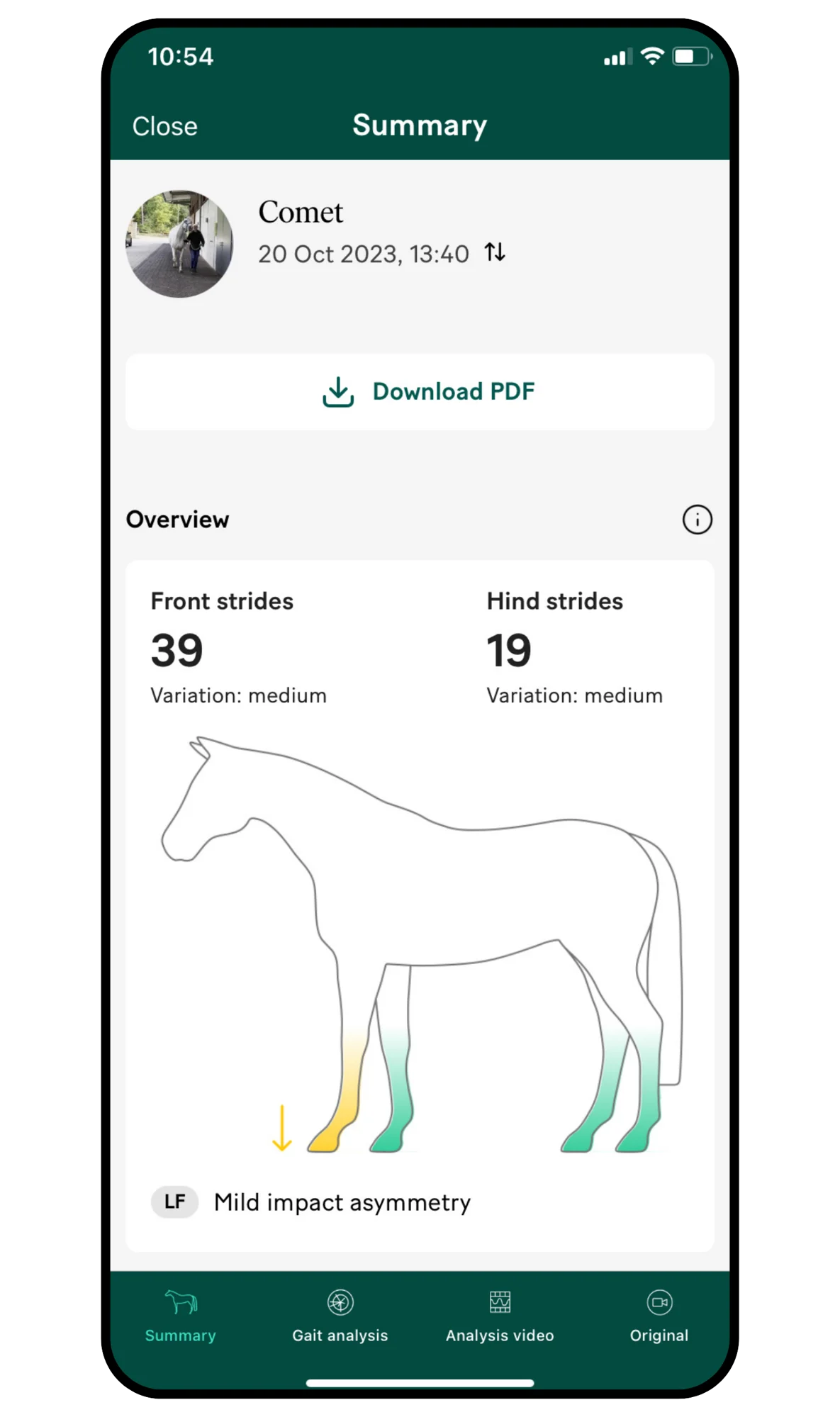
39 front limb strides have been included in this analysis. The variation noted between these strides is “medium”, which is acceptable. For the hind limbs, 19 strides were captured for analysis.
This overview gives you an indication of the quality of the data collected in your recording, based on the consistency of the trot and the number of strides included in the analysis. Around 20 analysed strides will give you a reliable measurement. The lower the variation between strides, the better.
Expect more front limb strides to be included as these are visible both when the horse is trotting away from the camera and when it faces the camera. The head will obscure some hindleg strides as the horse trots toward the camera.
A mild impact asymmetry was detected. No significant asymmetries in the right front or hindlimbs was detected.
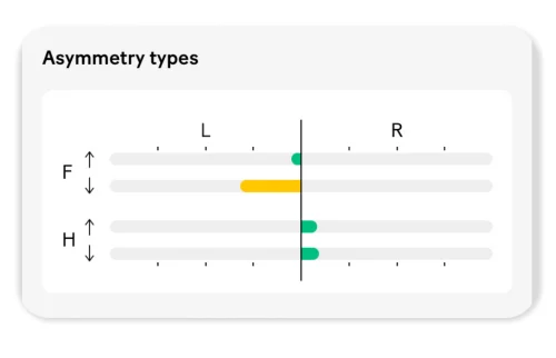
In the individual strides section, the impact asymmetry in the LF is quantified as 0,6, as visualised in the MinDiff. The MinDiff shows the difference between the two minima in the vertical position of the left and right half of the stride.
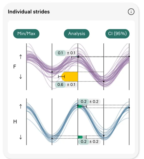
The lameness shows a consistent pattern in the left front; with strides clustered in the impact quadrant. The length of the lines in the circles indicates the severity of the asymmetry, which in this case is mild. The yellow line represents the mean value of the strides analysed. An outlier is seen in the hindlimbs, in the right-hand push-off quadrant.
The veterinarian administered pain blockers on the horse from distal to proximal. On the first examination day, Comet showed no improvement on a PDNB, nor on an intra-articular coffin joint block. The abaxial block revealed a very slight improvement, whereas blocking the ulnar nerve (ultrasound-guided) gave no improvement.
The final injection, administered at the fourth re-check some two and a half months later, was an intra-articular block of the elbow joint. After this block, a clear improvement could be seen. On the following page you’ll find the data output of the Sleip analysis performed following the final block.
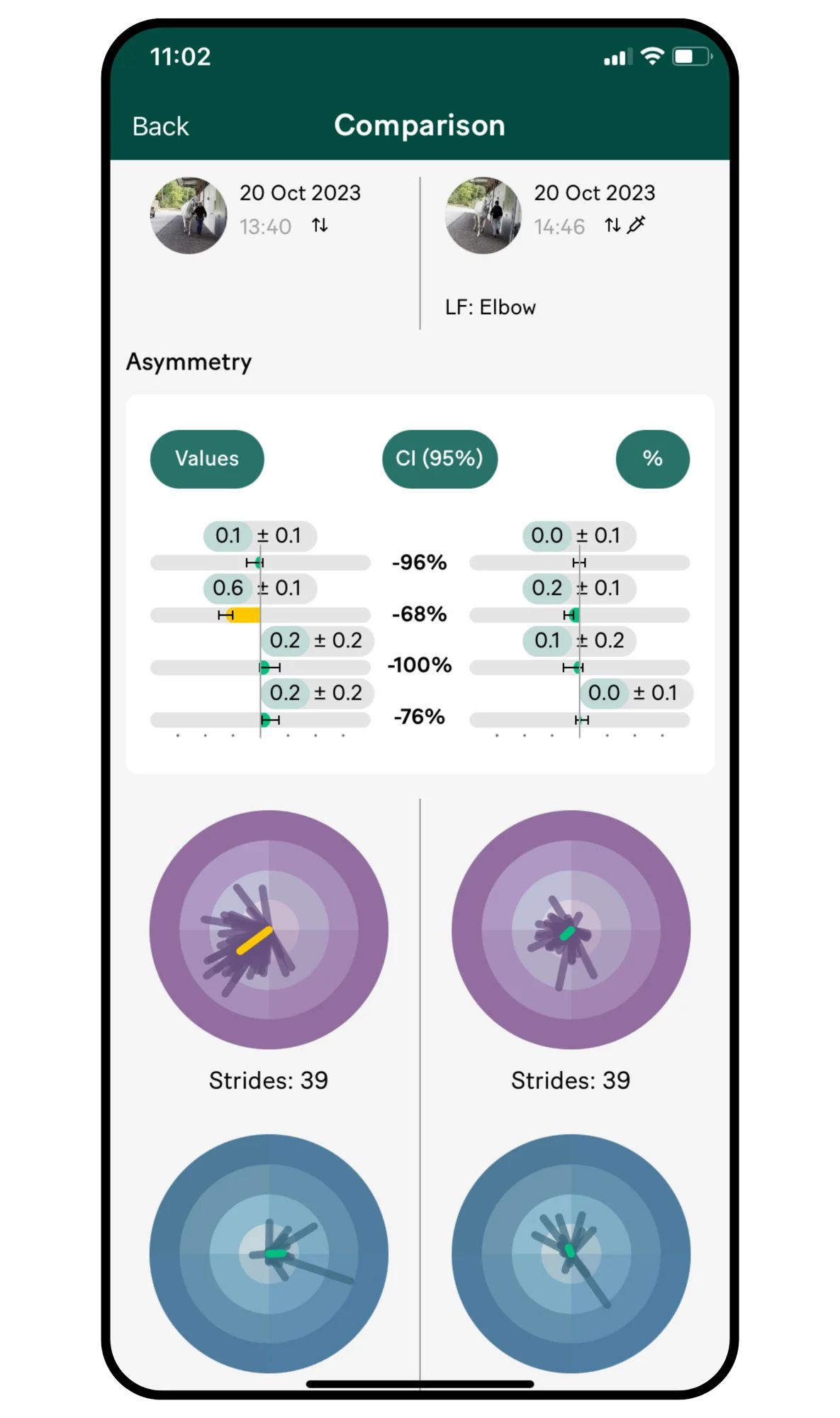
A significant reduction of the LF impact asymmetry can be seen.

The gait analysis plots and asymmetry data from the two recordings to be compared can be viewed side by side. The LF impact asymmetry decreased significantly, from 0.6 (0.1) to 0.2 (0.1), which is an improvement of 68 per cent.
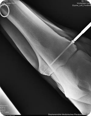
A radiographic examination revealed a bone cyst in the LF elbow.
Comet received surgical treatment of a subchondral lucency using a bone screw.