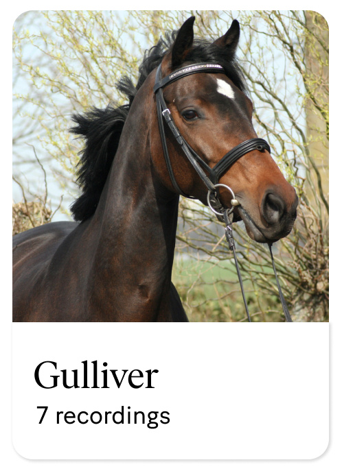
Breed: KWPN (Dutch Warmblood)
Gender: Gelding
Age: 9 years old
Discipline: Dressage
Training Level: Prix St. George (PSG)
Gulliver has been experiencing lameness in the left front leg for several months.
The horse has been hand-walked and Non-Steroidal Anti-Inflammatory Drugs (NSAIDs) have been administered.
There has been a minor improvement following the treatment.
The horse appears sound and coordinated at walk.
Lameness is evident in the left front limb with severity rated at 2-3 out of 5 on the lameness scale.
Increased severity on the left circle. On the longe, the horse displayed a lameness of 2 out of 5, on both hands. No significant change of the lameness after longing.
Visual assessment shows improvement after an abaxial block of the left front.
Examination surface: Hard
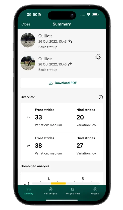
The overview gives you an indication of the quality of the data collected in your recording. It shows you the number of strides analysed and the variation in the stride pattern. The less variation between strides, the more certain the analysis. The more strides collected, the more certain the analysis. Around 20 analysed strides will give you a reliable measurement.
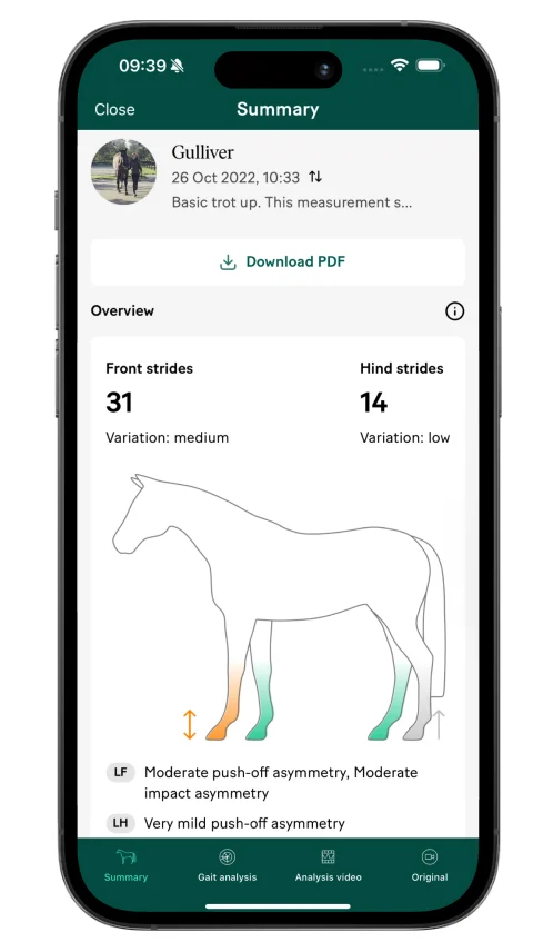
31 front limb strides have been included in this analysis. The variation noted between these strides is “medium”, which is acceptable. For the hind limbs, 14 strides have been captured in the recording and analysed, which is on the low side but acceptable as the variation in the stride pattern is “low”.
The analysis shows moderate asymmetries, both in the push-off and in the impact phase. The horse is not applying as much force or weight on the left front limb when it comes into contact with the ground (impact asymmetry) and also not using it as strongly to push off from the ground (push-off asymmetry), compared to the right front limb.
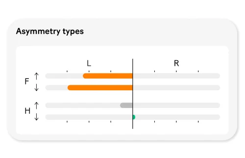
In the individual strides section, the asymmetry is quantified as 1.1 (with a standard deviation of 0.3) in the push-off phase and 1.5 (with a standard deviation of 0.3) in the impact phase. There is also a very mild asymmetry in the push-off phase in the left hind.
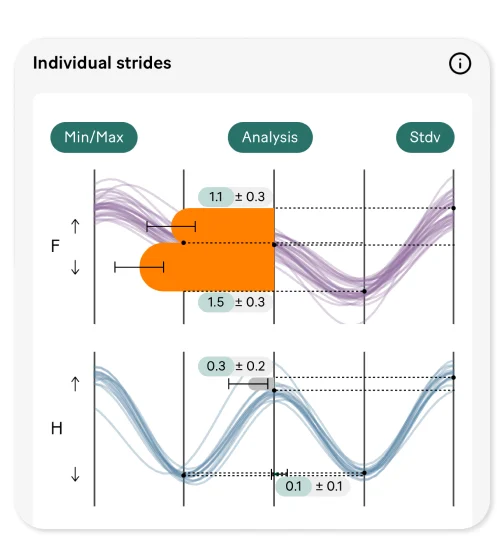
The lameness shows a very consistent pattern in the left front; all strides are clustered together, and the amplitude of the asymmetry (illustrated by the length of the lines) is moderate. In the hindlimbs there is also a very consistent asymmetry, but of a very low amplitude. One outlier is seen in the hindlimbs, in the right-hand push-off quadrant.

By assessing the slow-motion analysis video and data simultaneously, we can see that this outlier is caused by the first stride after turning the horse to trot away from the camera the second time.
Examination surface: Soft
Longeing data is collected in two separate recordings, one for each direction on the circle.
For the combined analysis, 33 front limb strides and 20 hind limb strides have been included for the left hand circle. For the right hand circle 38 and 27 strides have been included for the front and hind respectively. The variation noted between these strides is “medium”, which is acceptable given the number of strides included. For the hind limbs, fewer strides have been included but the variation between strides is low, making the total result reliable.
The combined analysis shown at the top shows a clear asymmetry on the left front.
The directional analysis shows that the asymmetry is more pronounced on the left circle in the push-off phase.
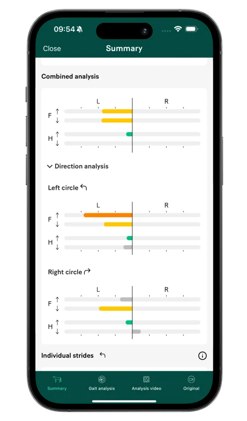
From this horse’s history, we know that it typically shows an inside impact asymmetry on the soft circle regardless of direction.
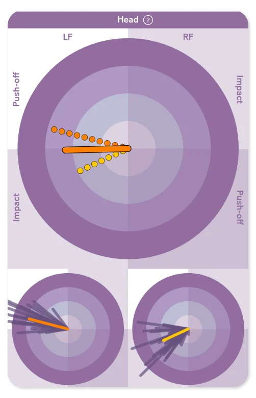
However, now the horse shows an impact asymmetry pointing towards the left on both hands. Additionally, there is a left-front oriented push-off asymmetry in both directions, although most prominent on the left circle.
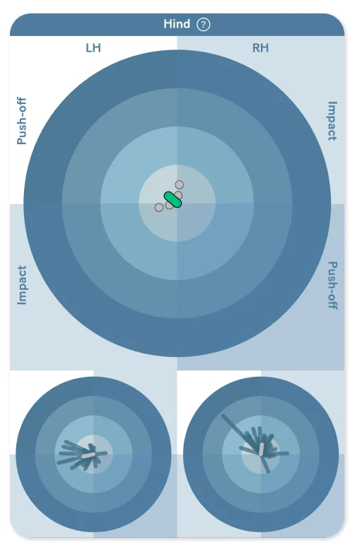
The gait analysis circle graphics for longeing analysis give a visual presentation of the combined analysis result in the larger circle at the top. The smaller circles at the bottom show you the analysis for the left-hand circle and the right-hand circle, respectively.
For the hindlimbs, there is a mild left hind asymmetry visible on the left circle. This mild asymmetry differs from the typical inside impact and outside push-off asymmetry observed in horses on the soft circle.
After the abaxial nerve block on the left front, the orange left front limb changed into a yellow limb and the the arrow up disappeared, meaning the asymmetry decreased and the push-off component disappeared. No further blocking was performed due to the clear improvement and the anamnesis of a chronic lameness.

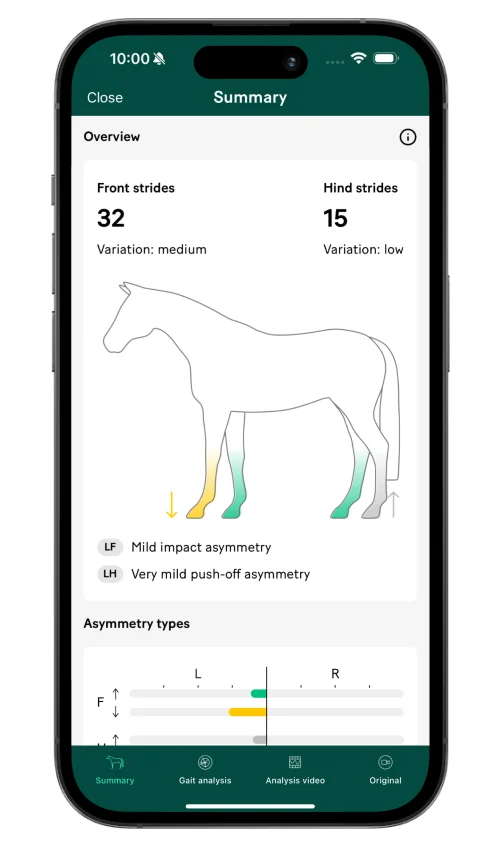
Quantitatively, there’s a significant improvement of 79% in the push-off and 62% in the impact component of the asymmetry in the left front after the abaxial block compared to baseline. The push-off asymmetry decreased from 1.1 to 0.2 (with a standard deviation of 0.2) and the impact asymmetry decreased from 1.5 to 0.6 (with a standard deviation of 0.3).
In the gait analysis circle graphics, we can clearly see that the stride-to-stride variation in the front increased while the amplitude of asymmetry decreased following the abaxial block. This is illustrated by the lines pointing in multiple directions instead of being clustered together closely. An increasing stride-to-stride variation is often seen when horses are trotted up following a positive block.

The Sleip measurements show a clear LF asymmetry with a significant decrease after the abaxial block, which is in line with the findings of the diagnostic imaging.
There is both skeletal damage (fracture of the coffin bone) and soft tissue damage (desmopathy of the medial collateral ligament of the coffin joint).
Fracture of the coffin bone, located medially
Severe sclerosis associated with diffuse fluid-based pathology in the medial ungual cartilage
Associated most likely with a fracture at its junction to the ipsilateral palmar process of the distal phalanx
Moderate diffuse fluid-based pathology at the medial palmar process of the distal phalanx
Mild to moderate diffuse desmopathy of the medial collateral ligament of the distal interphalangeal joint