Breed: Swedish Warmblood
Gender: Gelding
Age: 16 years old
Discipline: Dressage (leisure)
Coolman Q was first brought to Stockholms Hästklinik and DVM Sara Regårdh in May 2024, with an injury in the distal check ligament of the right forelimb (RF).
He was treated and spent the summer in a rehabilitation facility. In August 2024, during recovery, Coolman began showing signs of lameness in the LF. This prompted a full diagnostic work-up, including MRI and ultrasound scans.
The static examination showed no obvious clinical findings, no heat or pain upon palpation and negative on hoof-testers.
In motion, Coolman exhibited moderate lameness in the LF and mild lameness in the RH. Sleip substantiated and quantified these observations. Baseline recordings were performed on both the straight line and the longe. The following is the analysis video of the baseline measurement on the straight line.
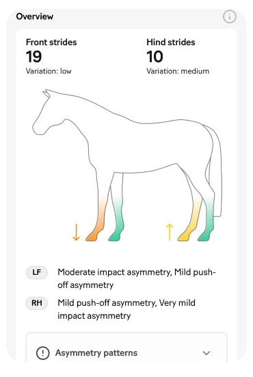
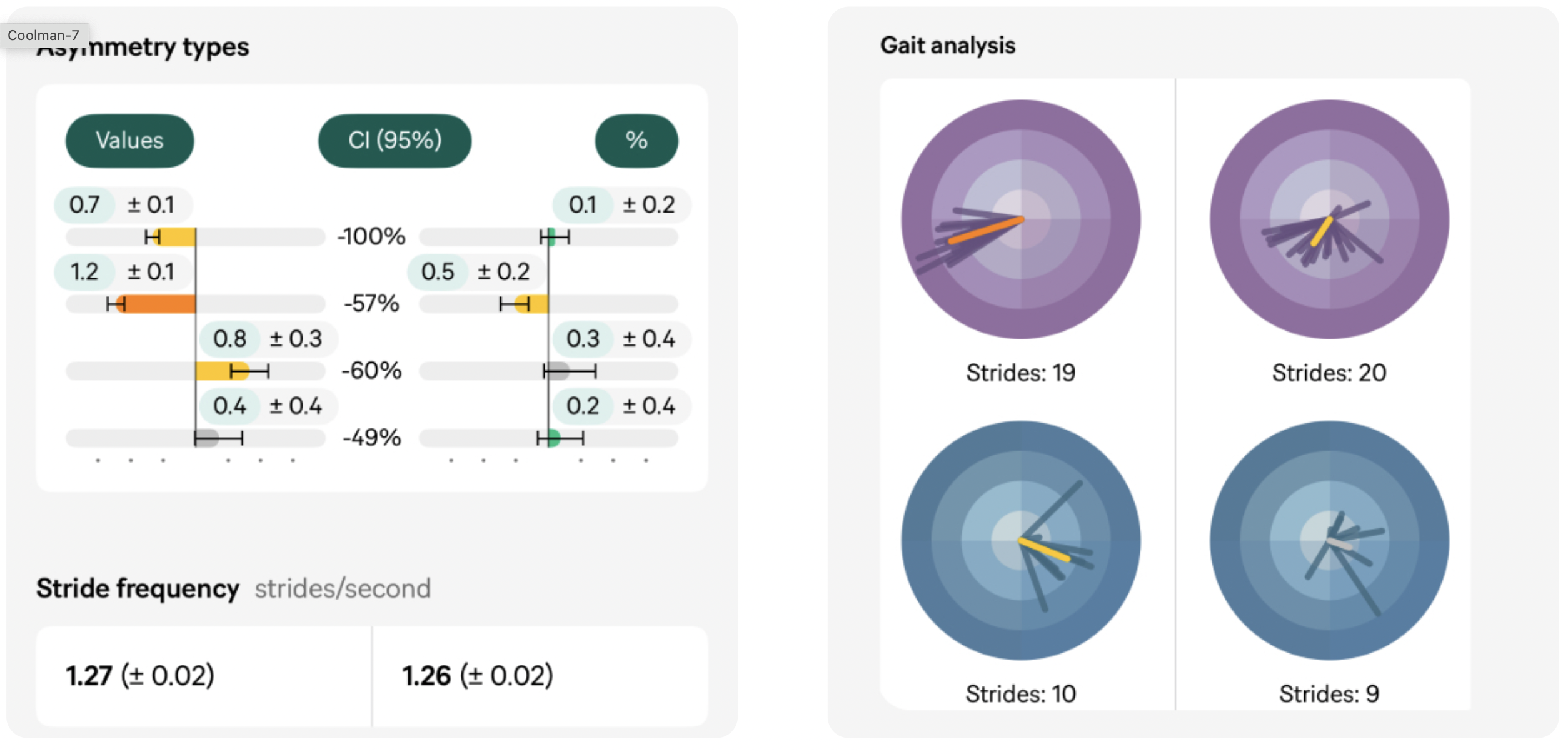
Summary overview of the analysis: Moderate asymmetry in the LF and a mild asymmetry in the RH.

A compensatory asymmetry pointing to the diagonal hind, and in some cases, the ipsilateral hindlimb, is a common response to a forelimb injury. Suspected compensatory patterns are flagged in the app.
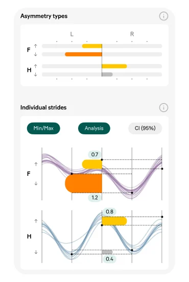
The asymmetry bars provide an additional overview of which limb the asymmetry originates from. Arrows pointing up indicate an issue in the push-off phase, and arrows pointing down indicate impact. The individual strides view provides the quantified measurements as well as a visual representation of each stride.
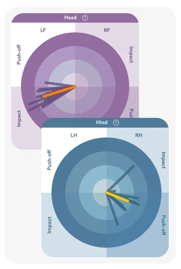
The vectors, where each line represents an individual stride, provide additional information about the consistency of the asymmetry. For the front limbs, the strides are clustered together, mainly pointing to the impact quadrant of the LF. The length of the lines illustrates the amplitude of the asymmetry, with the coloured line representing the mean. A milder asymmetry is noted in the push-off phase of the RH.
“At Stockholms Hästklinik, all patients are analysed with Sleip” — Sara Regårdh, DVM
Diagnostic analgesia of the palmar digital nerves (PDN) in the left forelimb resulted in a clear positive response.
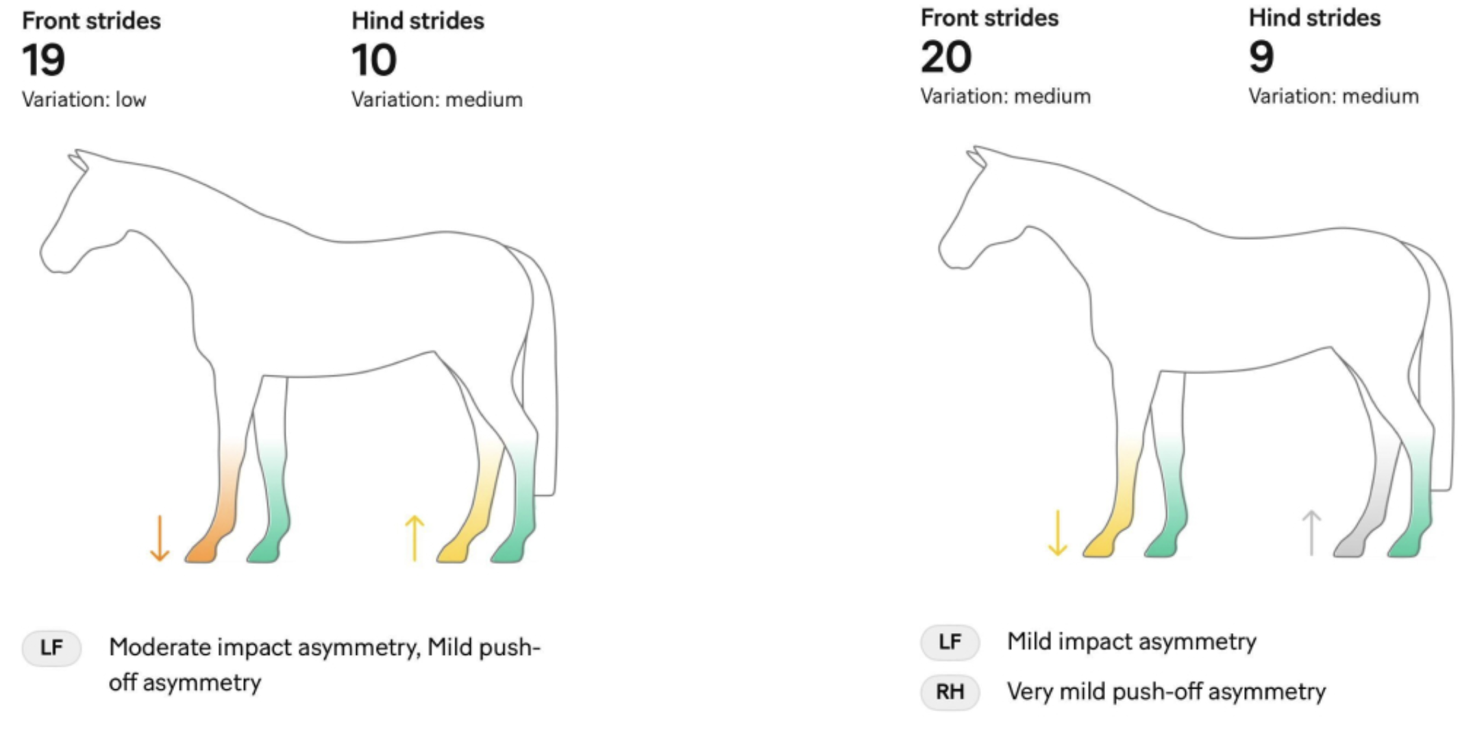
Comparing the baseline analysis with analysis after blocking using the comparison view provides a good indication of whether or not the block is positive. An improvement of around 50% is considered a positive block.
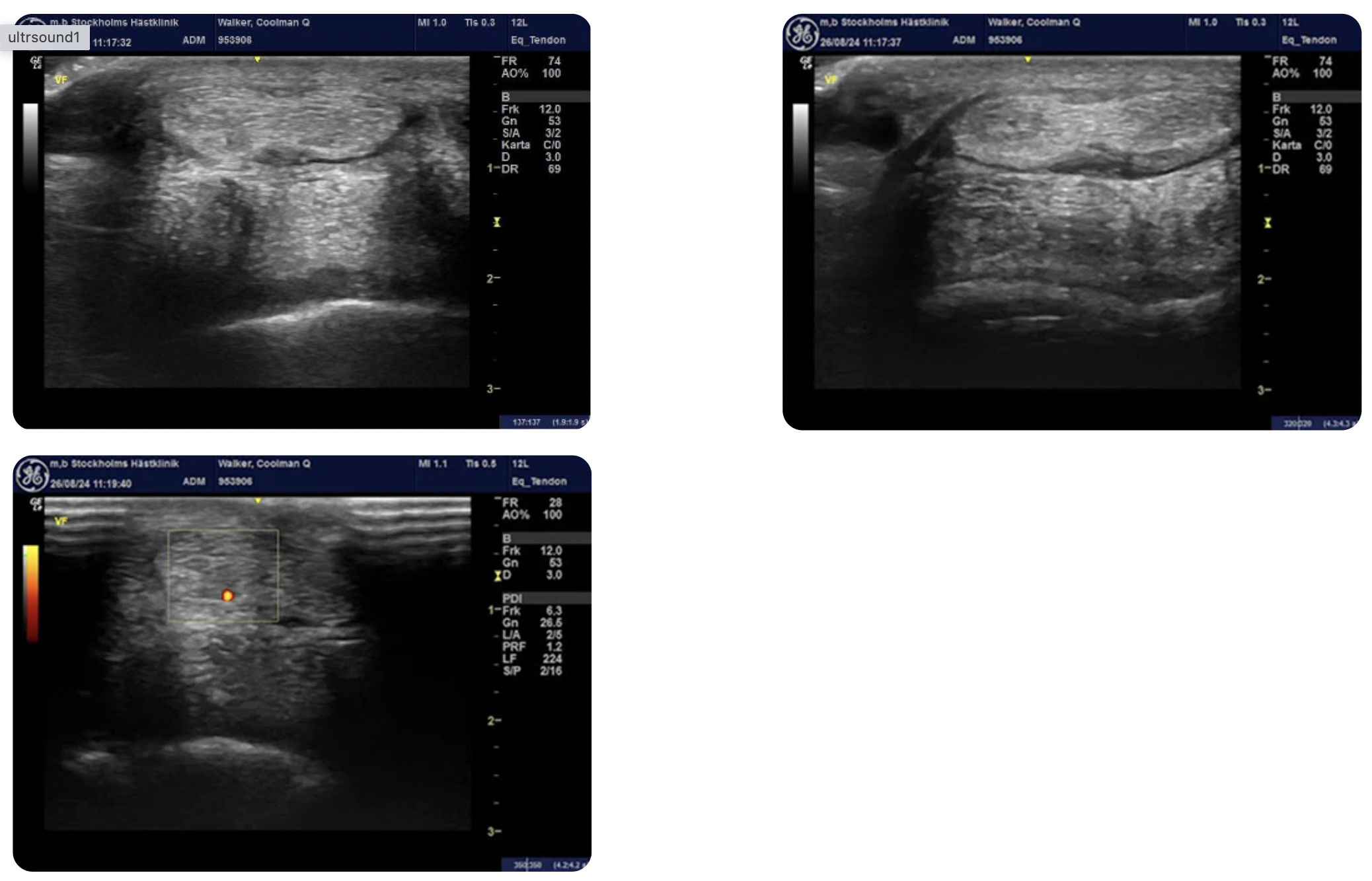
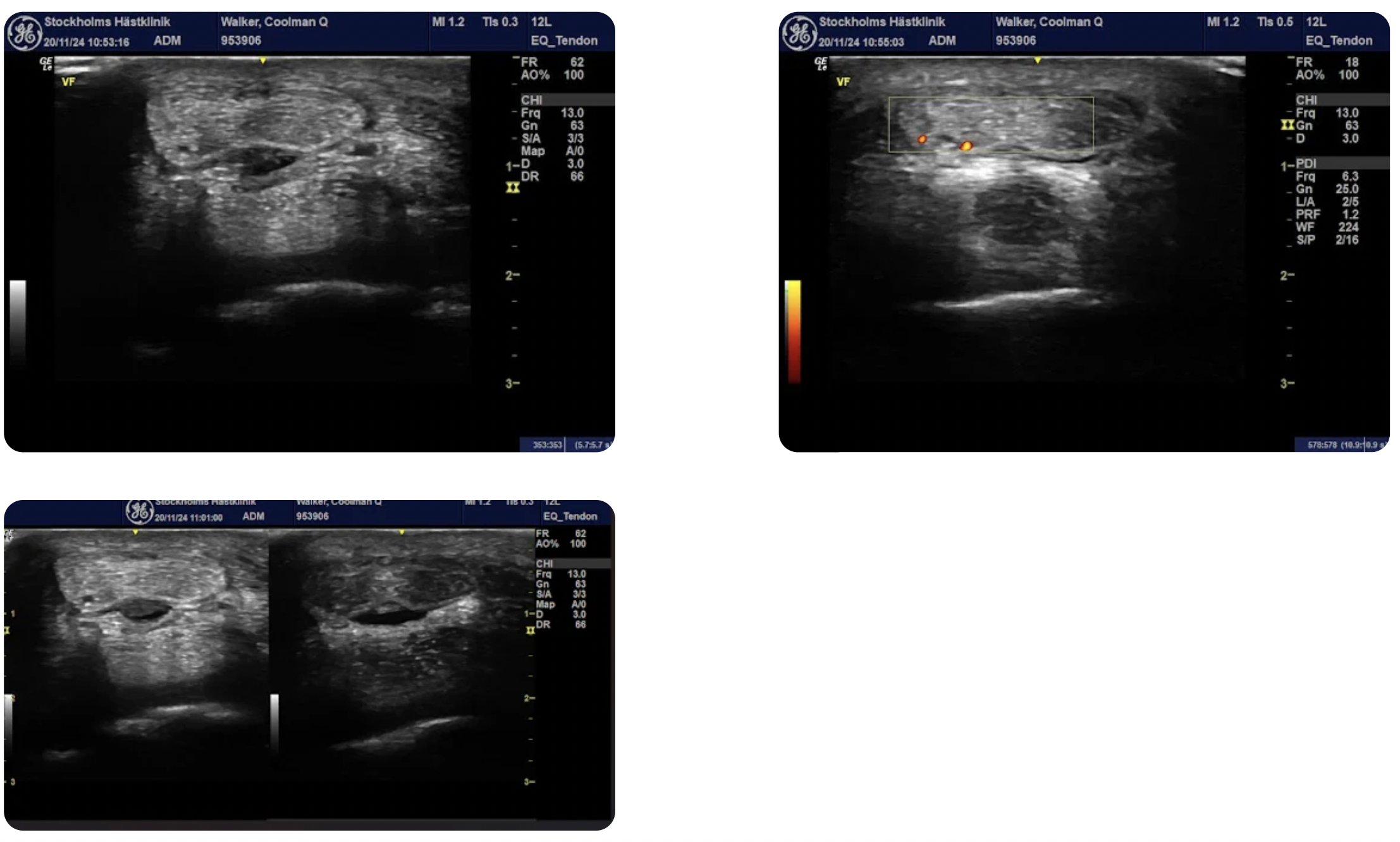
Upon examination with X-ray and ultrasound, Coolman Q was found to have a bulging medial lobe of the distal deep digital flexor tendon (DDFT) with a hypoechoic lesion on the palmar aspect at the level of P1. A positive Doppler signal was detected in the corresponding area as well as in the distal aspect of the DDFT, where the signal was more dorsally evident.
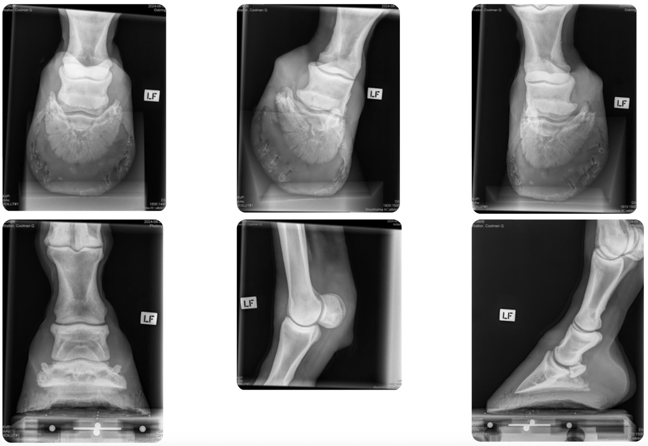
X-rays revealed mild synovial invaginations of the navicular bone, particularly on the medial aspect, along with slight remodeling of the palmar part of the navicular bone and a mild-to-moderate osteophyte on the proximal P1.The DDFT lesion was considered the most clinically relevant finding. Coolman Q was referred for an MRI of the left forelimb to assess the extent of the lesion.
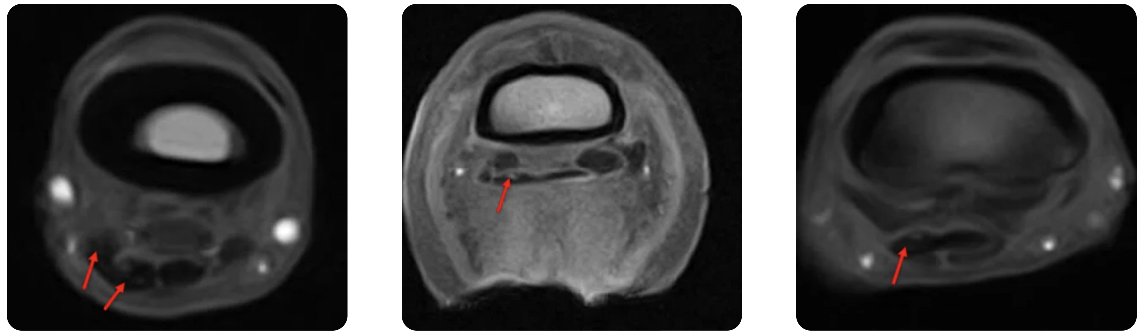
MRI confirmed an extensive lesion in the medial lobe of the DDFT, extending from distal P1 to its insertion on P3. Additional findings included moderate navicular bursitis in the left forelimb, an enlarged synovial fossa of the navicular bone with mild STIR hyperintensity—consistent with fluid-like material within the endosteal aspect of the navicular bone—mild injury to the medial branch of the superficial digital flexor tendon, mild-to-moderate synovial effusion of the distal interphalangeal joint, and sclerosis with moderate osteoarthritis of the fetlock joint.
Coolman Q has since undergone treatment with corrective shoeing, navicular bursa injections, shockwave therapy, and a controlled exercise program. He is progressing well in his rehabilitation.
Regular follow-ups and monitoring with Sleip help ensure that he is progressing well in his rehabilitation.
Sara Regårdh is one of four veterinarians at Stockholms Hästklinik, in Stockholm, Sweden. The clinic, established in 2019, specialises in equine locomotor health, dentistry, and diagnostic imaging, serving both sport and leisure horses.
Sara earned her veterinary degree from the University of Copenhagen in 2022. During her studies, she gained practical experience from the large animal teaching hospital in Denmarks and then completed externships in Sweden and the Netherlands. Her primary interests lie in lameness evaluations and diagnostic imaging. She is a member of the International Society of Equine Locomotor Pathology (ISELP).

